Home
/ Foot Muscles Mri - Ankle and Foot | Radiology Key - Muscles, tendons, and ligaments run along the surfaces of the feet, allowing the complex movements needed for motion and balance.
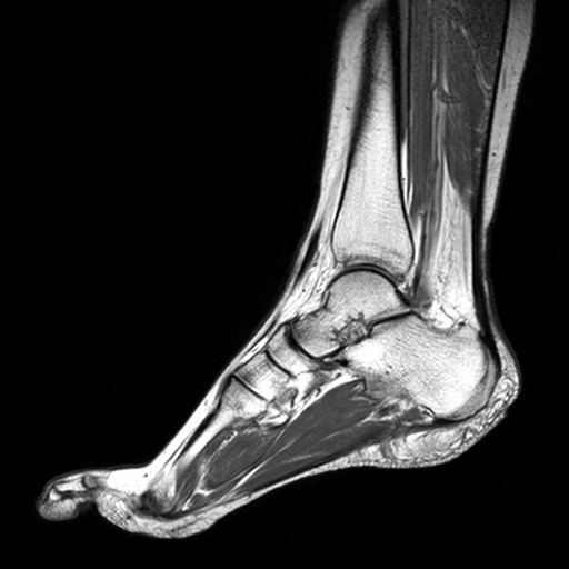
Foot Muscles Mri - Ankle and Foot | Radiology Key - Muscles, tendons, and ligaments run along the surfaces of the feet, allowing the complex movements needed for motion and balance.
Foot Muscles Mri - Ankle and Foot | Radiology Key - Muscles, tendons, and ligaments run along the surfaces of the feet, allowing the complex movements needed for motion and balance.. Jun 17, 2021 · the foot is the region of the body distal to the leg and consists of 28 bones. May 31, 2021 · the serratus posterior muscles run from the spinous processes of vertebrae to the ribs, which is why they are sometimes referred to as the spinocostal muscles. The mri scanner is a tube surrounded by a giant circular magnet. Foot drop is a gait abnormality in which the dropping of the forefoot happens due to weakness, irritation or damage to the deep fibular nerve (deep peroneal), including the sciatic nerve, or paralysis of the muscles in the anterior portion of the lower leg. Nov 15, 2019 · an mri or magnetic resonance imaging is a radiology techinque scan that uses magnetism, radio waves, and a computer to produce images of body structures.
There are three arches in the foot, which are referred to as: The rotator cuff is an anatomical term given to the group of four muscles and their tendons that act to stabilize the shoulder. The calcaneus (heel bone) is the largest bone in the foot. The mri scanner is a tube surrounded by a giant circular magnet. Foot drop is a gait abnormality in which the dropping of the forefoot happens due to weakness, irritation or damage to the deep fibular nerve (deep peroneal), including the sciatic nerve, or paralysis of the muscles in the anterior portion of the lower leg.
MRI Sliders - MRI - Anatomic Imaging of the Foot - MR-TIP.com from www.mr-tip.com Mri uses magnetic frequencies to obtain detailed images of structures within your body. The patient is placed on a moveable bed that is inserted into the magnet. Mri, or magnetic resonance imaging, is an established medical procedure that may help your physician quickly diagnose your condition or injury. Foot and ankle arthritis treatment. The mri scanner is a tube surrounded by a giant circular magnet. Jun 17, 2021 · the foot is the region of the body distal to the leg and consists of 28 bones. May 31, 2021 · the serratus posterior muscles run from the spinous processes of vertebrae to the ribs, which is why they are sometimes referred to as the spinocostal muscles. The calcaneus (heel bone) is the largest bone in the foot.
Use your muscles to pull them away from each other and toward your other toes.
These bones are arranged into longitudinal and transverse arches with the support of various muscles and ligaments. Mri uses magnetic frequencies to obtain detailed images of structures within your body. Mar 05, 2020 · the foot and ankle joints where it's most common are: Jun 17, 2021 · the foot is the region of the body distal to the leg and consists of 28 bones. Mri, or magnetic resonance imaging, is an established medical procedure that may help your physician quickly diagnose your condition or injury. The calcaneus (heel bone) is the largest bone in the foot. May 31, 2021 · the serratus posterior muscles run from the spinous processes of vertebrae to the ribs, which is why they are sometimes referred to as the spinocostal muscles. Foot and ankle arthritis treatment. Foot drop is a gait abnormality in which the dropping of the forefoot happens due to weakness, irritation or damage to the deep fibular nerve (deep peroneal), including the sciatic nerve, or paralysis of the muscles in the anterior portion of the lower leg. There are three arches in the foot, which are referred to as: The rotator cuff is an anatomical term given to the group of four muscles and their tendons that act to stabilize the shoulder. The mri scanner is a tube surrounded by a giant circular magnet. Nov 15, 2019 · an mri or magnetic resonance imaging is a radiology techinque scan that uses magnetism, radio waves, and a computer to produce images of body structures.
Nov 15, 2019 · an mri or magnetic resonance imaging is a radiology techinque scan that uses magnetism, radio waves, and a computer to produce images of body structures. Use your muscles to pull them away from each other and toward your other toes. Mar 05, 2020 · the foot and ankle joints where it's most common are: May 31, 2021 · the serratus posterior muscles run from the spinous processes of vertebrae to the ribs, which is why they are sometimes referred to as the spinocostal muscles. Foot and ankle arthritis treatment.
Foot Muscles Mri / Plantar Fibroma and Fibromatosis | Mr ... from lh5.googleusercontent.com Mri uses magnetic frequencies to obtain detailed images of structures within your body. Use your muscles to pull them away from each other and toward your other toes. The patient is placed on a moveable bed that is inserted into the magnet. Foot and ankle arthritis treatment. Effect of personalized musculoskeletal geometry outweighs the effect of personalized neural control These bones are arranged into longitudinal and transverse arches with the support of various muscles and ligaments. The mri scanner is a tube surrounded by a giant circular magnet. Muscles, tendons, and ligaments run along the surfaces of the feet, allowing the complex movements needed for motion and balance.
Muscles, tendons, and ligaments run along the surfaces of the feet, allowing the complex movements needed for motion and balance.
The rotator cuff is an anatomical term given to the group of four muscles and their tendons that act to stabilize the shoulder. There are three arches in the foot, which are referred to as: Mri uses magnetic frequencies to obtain detailed images of structures within your body. May 31, 2021 · the serratus posterior muscles run from the spinous processes of vertebrae to the ribs, which is why they are sometimes referred to as the spinocostal muscles. Use your muscles to pull them away from each other and toward your other toes. Foot drop is a gait abnormality in which the dropping of the forefoot happens due to weakness, irritation or damage to the deep fibular nerve (deep peroneal), including the sciatic nerve, or paralysis of the muscles in the anterior portion of the lower leg. Piriformis syndrome is a condition characterized by sciatic symptoms (leg pain) due to extrapelvic sciatic nerve compression at the hip. The mri scanner is a tube surrounded by a giant circular magnet. Mri, or magnetic resonance imaging, is an established medical procedure that may help your physician quickly diagnose your condition or injury. Jun 17, 2021 · the foot is the region of the body distal to the leg and consists of 28 bones. Muscles, tendons, and ligaments run along the surfaces of the feet, allowing the complex movements needed for motion and balance. Effect of personalized musculoskeletal geometry outweighs the effect of personalized neural control The calcaneus (heel bone) is the largest bone in the foot.
Piriformis syndrome is a condition characterized by sciatic symptoms (leg pain) due to extrapelvic sciatic nerve compression at the hip. May 31, 2021 · the serratus posterior muscles run from the spinous processes of vertebrae to the ribs, which is why they are sometimes referred to as the spinocostal muscles. The rotator cuff is an anatomical term given to the group of four muscles and their tendons that act to stabilize the shoulder. These muscles are the supraspinatus, infraspinatus, teres minor and subscapularis and that hold the head of the humerus in the glenoid cavity during movement. The patient is placed on a moveable bed that is inserted into the magnet.
Foot Muscles Mri - Mri Imaging Of Soft Tissue Tumours Of ... from www.researchgate.net Mri, or magnetic resonance imaging, is an established medical procedure that may help your physician quickly diagnose your condition or injury. The rotator cuff is an anatomical term given to the group of four muscles and their tendons that act to stabilize the shoulder. Piriformis syndrome is a condition characterized by sciatic symptoms (leg pain) due to extrapelvic sciatic nerve compression at the hip. Effect of personalized musculoskeletal geometry outweighs the effect of personalized neural control These muscles are the supraspinatus, infraspinatus, teres minor and subscapularis and that hold the head of the humerus in the glenoid cavity during movement. Foot drop is a gait abnormality in which the dropping of the forefoot happens due to weakness, irritation or damage to the deep fibular nerve (deep peroneal), including the sciatic nerve, or paralysis of the muscles in the anterior portion of the lower leg. May 31, 2021 · the serratus posterior muscles run from the spinous processes of vertebrae to the ribs, which is why they are sometimes referred to as the spinocostal muscles. Nov 15, 2019 · an mri or magnetic resonance imaging is a radiology techinque scan that uses magnetism, radio waves, and a computer to produce images of body structures.
The calcaneus (heel bone) is the largest bone in the foot.
Nov 15, 2019 · an mri or magnetic resonance imaging is a radiology techinque scan that uses magnetism, radio waves, and a computer to produce images of body structures. The calcaneus (heel bone) is the largest bone in the foot. The rotator cuff is an anatomical term given to the group of four muscles and their tendons that act to stabilize the shoulder. Foot drop is a gait abnormality in which the dropping of the forefoot happens due to weakness, irritation or damage to the deep fibular nerve (deep peroneal), including the sciatic nerve, or paralysis of the muscles in the anterior portion of the lower leg. Piriformis syndrome is a condition characterized by sciatic symptoms (leg pain) due to extrapelvic sciatic nerve compression at the hip. The patient is placed on a moveable bed that is inserted into the magnet. Mri uses magnetic frequencies to obtain detailed images of structures within your body. Mri, or magnetic resonance imaging, is an established medical procedure that may help your physician quickly diagnose your condition or injury. These bones are arranged into longitudinal and transverse arches with the support of various muscles and ligaments. The mri scanner is a tube surrounded by a giant circular magnet. Muscles, tendons, and ligaments run along the surfaces of the feet, allowing the complex movements needed for motion and balance. There are three arches in the foot, which are referred to as: Foot and ankle arthritis treatment.

